|
|
|
HCR RNA–FISH v3.0
State-of-the-art RNA imaging:
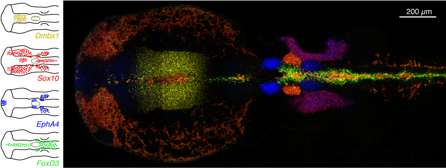
- Straightforward multiplexing with 1–step HCR signal amplification for up to 10 targets simultaneously
- Analog RNA relative quantitation with subcellular resolution (qHCR imaging)
- Digital RNA absolute quantitation with single–molecule resolution (dHCR imaging)
- Automatic background suppression throughout the protocol for dramatically enhanced performance (signal–to–background, qHCR precision, dHCR fidelity) and ease–of–use (no probe set optimization)
- Deep sample penetration (e.g., entire adult mouse brain)
- High signal–to–background in highly autofluorescent samples
- Compatible with HCR IF and HCR PPI
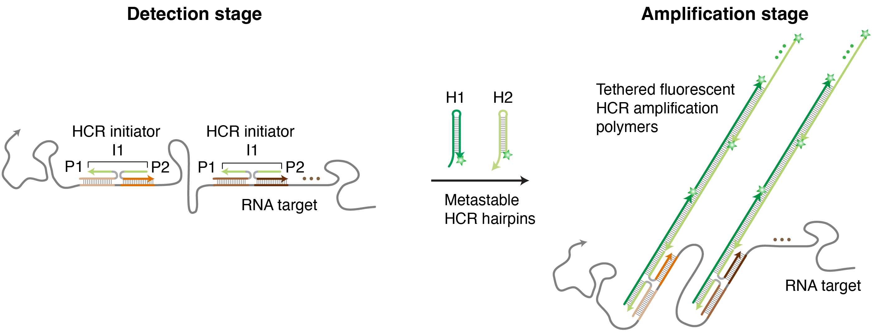
HCR RNA-FISH (v3.0): split-initiator probes (P1,P2) colocalize a full HCR initiator I1 to trigger HCR signal amplification only if they bind specifically to the target RNA ( Choi et al., 2018).
|
|